1. Positron Emission Tomography (PET) Scans: How It Works, How do you Prepare? Risks, Technology Used
Positron emission tomography (PET) is a nuclear imaging tool that produces a 3D map of the functional areas of the body including the lungs, heart, chest or abdominal area as well as other internal organs.
A PET scan can help the doctor see how the tissues and organs in your body are functioning. Unlike Magnetic Resonance Imaging (MRI), Computed Tomography (CT) scans or X-Rays that are designed to show visuals of the structural details of your organs, a PET scan was designed to show chemical activities within certain organs and tissues in your body.
This chemical activity occurring in your organs may indicate areas where tumors reside, and that other imaging techniques have failed to detect. Thus, a Positron Emission Tomography (PET) scan is very useful in examining a variety of conditions including heart disease, body inflammation caused by asbestos fibers, infections and other neurological diseases.
2. Magnetic Resonance Imaging (MRI) Scans for Mesothelioma Lung Cancer: How It Works, Magnetic Resonance? Superconducting Magnets & Precautions
Magnetic Resonance Imaging (MRI) is a medical imaging technique used to take pictures of the lungs, structures of the body and organs. It can provide detailed visuals of the body in any plane.
The advantage of using MRI over Computed Tomography (CT) scans is that MRI scans provide greater contrast between different tissues of the body making it easier to detect malignant cancerous cells & tumors.
While CT scans use ionizing radiation or X-rays to acquire images of the lungs, MRI scans use non-ionizing radio frequency (RF) signals to output images of internal organs of the body.
To determine extent & development of tumors in the lungs, Magnetic Resonance Imaging (MRI) scans use strong magnets & radio waves from which the energy released is formed in a pattern.
A specialized computer translates these patterns of radio waves emitted from the tissues into very detailed images of the body.
Not only does this produce cross sectional slices of the lungs or the body, it can also output slices that are parallel with the length of the body.
MRI scans are also used to visualize the diaphragm (thin muscle at the bottom of the lungs that helps the body respirate) where malignant mesothelioma tumors could easily spread to.
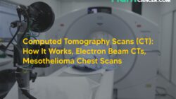
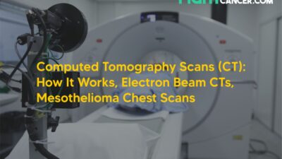
3. Computed Tomography Scans (CT): How It Works, Electron Beam CTs, Mesothelioma Chest Scans
Computed Tomography (CT) is a form of medical imaging that uses the concept of Tomography to deliver x-ray images in 2 dimensional cross–sectional formats.
Once the mesothelioma doctor performs x-rays on the patient’s lungs and if they show pleura or lung abnormalities, the doctor may ask him to undergo a CT scan to deliver more precise results.
The patient will be asked to lie on a couch and a scanning machine will be put over the head. The machine will rotate 180 degrees emitting thin x-ray beams at multiple points.
Crystals at opposite ends of the beam will record the absorption rates of varying thickness levels of tissues and your bones.
The machine will then turn these x-ray images into a detailed picture allowing the doctor to make conclusions & recommendations. Here are some of the advantages of using computed tomography scans over x-rays:
- CT scans allow the radiologist to see detailed views of the lungs & the pleura
- CT scans help determine the location, extent & size of tumor masses residing in the lungs more accurately than x-rays
- CT scans can reveal thickening of the pleura by examining the absorption rates of varying thickness levels of tissues
- CT scans can also indicate lung cancer beyond the pleura within the chest wall or lymph nodes
- CT scans can help evaluate the conditions of the lungs
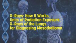
4. X-Rays – How It Works, Units of Radiation Exposure, X-Rays of the Lungs for Diagnosing Mesothelioma
X-ray is a form of electro-magnetic radiation with a wavelength of 10 to 0.01 nanometers; much like gamma rays but shorter than UV rays. X-rays contain high energy radiation exposure because they have an extremely short wavelength and high frequencies.
Just like Computed Tomography (CT) scans, X-Rays use ionizing radiation to create radio waves to create visuals of different organs of the body including the lungs.
Once the x-ray machine aims at the part of the body that is to be visualized such as the lungs, it will emit a small burst of radiation that will pass through the skin and record image of internal organs of the body on a photographic film or a special image recording plate. Different organs of the body will absorb the x ray radiation in different ways.
For instance dense bones will absorb almost all of the radiation while soft tissues such as muscles, fats & other organs will allow more of the x-rays to pass through them.
Due to this, bones appear white on x-rays while soft tissues are presented in shades of grey and black. X-Rays are also very similar to visible light rays where electromagnetic energy is carried by particles known as photons.
The difference between x-rays and visible light rays is the energy levels of individual photons, also known as the ‘Wavelength.
dr. Khadijah