Computed Tomography (CT) is a form of medical imaging that uses the concept of Tomography to deliver x-ray images in 2 dimensional cross–sectional formats.
Once the mesothelioma doctor performs x-rays on the patient’s lungs and if they show pleura or lung abnormalities, the doctor may ask him to undergo a CT scan to deliver more precise results.
The patient will be asked to lie on a couch and a scanning machine will be put over the head. The machine will rotate 180 degrees emitting thin x-ray beams at multiple points.
Crystals at opposite ends of the beam will record the absorption rates of varying thickness levels of tissues and your bones.
The machine will then turn these x-ray images into a detailed picture allowing the doctor to make conclusions & recommendations. Here are some of the advantages of using computed tomography scans over x-rays:
- CT scans allow the radiologist to see detailed views of the lungs & the pleura
- CT scans help determine the location, extent & size of tumor masses residing in the lungs more accurately than x-rays
- CT scans can reveal thickening of the pleura by examining the absorption rates of varying thickness levels of tissues
- CT scans can also indicate lung cancer beyond the pleura within the chest wall or lymph nodes
- CT scans can help evaluate the conditions of the lungs
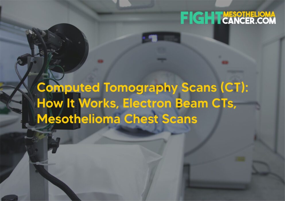
How CT Scans Work
The machine on the left is known as a ‘Gantry.’ It is a doughnut shaped structure on which the patient will lie and contains x-ray tubes and special electronic detectors that make the CT scan work.
X-ray data is obtained using an x-ray tube that rotates around the patient. X-ray sensors are placed on each side of the gantry (shown as ‘detectors’ in this picture) and they use the ionization principle to capture scans of the patient’s lungs.
A specialized form of computed axial tomography (CAT or CT) is the Electron beam tomography (EBT) where the x-ray tube is not mechanically spun in order to rotate the x-ray photons.
The x-ray source point moves around the circle where the patient is lying and captures images. Electron beam tomography (EBT) can capture complete view of the heart structures with every single heart beat; this process is very fast and produces images from a wide variety of angles allowing doctors to make accurate diagnosis.
The main advantage of using EBT tomographic CT machines over conventional CT scanners is that because the X-ray source point moves electronically and not mechanically, it can capture many images at rapid speeds.
Since the heart never stops moving and the lungs and arteries move several times their diameter for each heartbeat, scientists thought it was better to build a device that could be just as fast as the arteries, and thus Electron Beam Tomography was born.
The most advanced EBT scanner can perform a sweep in 0.025 seconds. On the other hand, the fastest conventional X-ray tube design scanners take about 0.33 seconds to perform 1 sweep (capture 1 image).
How do you prepare for a CT scan? The radiologist will ask you to wear an examination gown and all jewellery from the waist to the top of the body should be removed. Jewellery can obscure the CT scanner from taking proper images of the chest, and thus should be removed.
Also, if you are pregnant whilst taking a CT scan, inform your doctor right away. This is because radiation exposure from the scan could negatively impact the baby and the patient is advised NOT to take the CT scan while pregnant.
Radiation Exposure
CT scans are classified as moderate to high radiation diagnostic tools due to the complexity in taking images of the heart with every heartbeat.
The amount of radiation exposure depends on many factors including patient build, volume scanned (# of sweeps), type of scan sequences and desired resolution/image quality.
It is estimated that radiation exposure from current CT scan use could cause as many as 1 in 50 future cases of Cancer.
dr. Khadijah